Home
Uncategories
Abdominal Anatomy Aorta - Abdominal Aortic Aneurysm Health Information Bupa Uk - It thus follows the curvature of the lumbar vertebrae, that is, convex anteriorly.
Abdominal Anatomy Aorta - Abdominal Aortic Aneurysm Health Information Bupa Uk - It thus follows the curvature of the lumbar vertebrae, that is, convex anteriorly.
Abdominal Anatomy Aorta - Abdominal Aortic Aneurysm Health Information Bupa Uk - It thus follows the curvature of the lumbar vertebrae, that is, convex anteriorly.. The aorta runs from your heart through the center of your chest and abdomen. Pain is the most common symptom of an abdominal aortic aneurysm. The abdominal aorta has three distinct tissue layers: It is approximately 13cm long and ends at the level of the l4 vertebra. The abdominal aorta courses through the retroperitoneal space of the abdominal cavity.
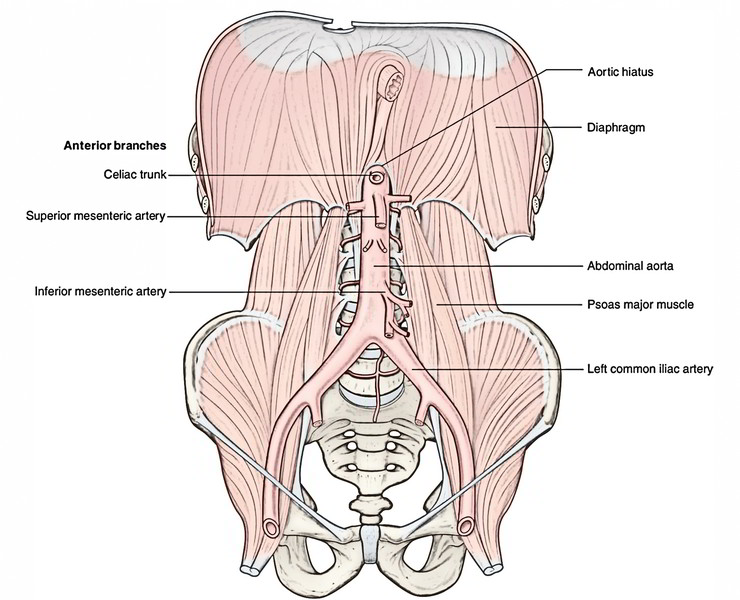
It travels down the posterior wall of the abdomen, anterior to the vertebral column. Abdominal aorta the abdominal aorta runs from the diaphragm and ends just above the pelvis, where it divides into the iliac arteries. At the renal hila, its mean. The abdominal aorta courses through the retroperitoneal space of the abdominal cavity. Abdominal aorta anatomy the portion of the aorta that begins below the diaphragm, extends to the bifurcation of the iliac arteries, and supplies blood to the abdominal viscera, pelvic organs and legs branches inferior phrenic, lumbar, celiac trunk, superior mesenteric, inferior mesenteric, middle adrenal, renal, testicular and ovarian arteries
Abdominal Aorta Branches Youtube from i.ytimg.com The abdominal aorta has three distinct tissue layers: • the abdominal aorta begins at the diaphragm, splitting to become the paired iliac arteries in the lower abdomen. Risk of rupture is proportional to the size of. The abdominal aorta is the final section of the aorta, the largest artery in the body. Anteriorly, the abdominal aorta is related to the organs of the abdominal cavity. At this level, the aorta terminates by bifurcating into the right and left common iliac arteries that supply the lower body. It is a continuation of the thoracic aorta. It begins at the diaphragm, and runs down to the point where it ends (by splitting in two to form the common iliac arteries).
It is a continuation of the thoracic aorta.
Abdominal aorta the abdominal aorta continues from the thoracic aorta as it passes posterior to the median arcuate ligament and between the crura of the diaphragm (aortic hiatus), in front of the body of the t12 vertebra and then descends slightly to the left of midline. At this level, the aorta terminates by bifurcating into the right and left common iliac arteries that supply the lower body. The media comprises smooth muscle cells surrounded by. At the weak portion, the aneurysm bulges and poses a. This video covers the anatomy and branches of the abdominal aorta. Abdominal aorta anatomy the portion of the aorta that begins below the diaphragm, extends to the bifurcation of the iliac arteries, and supplies blood to the abdominal viscera, pelvic organs and legs branches inferior phrenic, lumbar, celiac trunk, superior mesenteric, inferior mesenteric, middle adrenal, renal, testicular and ovarian arteries Most aneurysms grow slowly (~10%/year) without causing symptoms, and most are found incidentally. It is the continuation of the thoracic aorta after it enters the abdomen via the aortic hiatus of the diaphragm. An ultrasound image of an abdominal aortic aneurysm is shown in the upper right corner. The enlarged area in the lower part of the aorta is an abdominal aortic aneurysm. It is approximately 13cm long and ends at the level of the l4 vertebra. The abdominal aorta begins at the diaphragmatic hiatus. The aorta is the largest blood vessel in the body.
At the weak portion, the aneurysm bulges and poses a. 531) begins at the aortic hiatus of the diaphragm, in front of the lower border of the body of the last thoracic vertebra, and, descending in front of the vertebral column, ends on the body of the fourth lumbar vertebra, commonly a little to the left of the middle line, (* 103 by dividing into the two common iliac arteries. It is a continuation of the thoracic aorta. The abdominal arteries arise from the abdominal aorta and are comprised of three groups of arteries: For even more information about the aorta, check out our free article on kenhub:
Easy Notes On Abdominal Aorta Learn In Just 3 Minutes Earth S Lab from www.earthslab.com The abdominal aorta courses through the retroperitoneal space of the abdominal cavity. The normal caliber of the abdominal aorta increases with age; Abdominal aorta (page 223) dr. • the abdominal aorta begins at the diaphragm, splitting to become the paired iliac arteries in the lower abdomen. Aortas or aortae 4) is the main blood vessel in the abdominal cavity that transmits oxygenated blood from the thoracic cavity to the organs within the abdomen and to the lower limbs. The abdominal aorta in a nutshell the abdominal aorta is a continuation of the descending thoracic aorta. The abdominal aorta supplies oxygenated blood to all of the abdominal and pelvic organs and the legs. 531) begins at the aortic hiatus of the diaphragm, in front of the lower border of the body of the last thoracic vertebra, and, descending in front of the vertebral column, ends on the body of the fourth lumbar vertebra, commonly a little to the left of the middle line, (* 103 by dividing into the two common iliac arteries.
531) begins at the aortic hiatus of the diaphragm, in front of the lower border of the body of the last thoracic vertebra, and, descending in front of the vertebral column, ends on the body of the fourth lumbar vertebra, commonly a little to the left of the middle line, (* 103 by dividing into the two common iliac arteries.
The abdominal aorta begins at the diaphragmatic hiatus. Pain is the most common symptom of an abdominal aortic aneurysm. For even more information about the aorta, check out our free article on kenhub: It is an artery, meaning that it carries blood away from the heart. Abdominal aorta the abdominal aorta runs from the diaphragm and ends just above the pelvis, where it divides into the iliac arteries. Aortas or aortae 4) is the main blood vessel in the abdominal cavity that transmits oxygenated blood from the thoracic cavity to the organs within the abdomen and to the lower limbs. The cause is multifactorial, but atherosclerosis is often involved. It is a continuation of the thoracic aorta. Abdominal ultrasound of an abdominal aortic aneurysm. The unpaired visceral arteries supply the gastrointestinal (gi) tract, spleen, pancreas, gallbladder, and liver and are made up of the celiac trunk, superior mesenteric. The abdominal aorta enters the abdomen through the diaphragm at the level of the twelfth thoracic vertebre and continues to just below the umbilical area, where it splits into the right and left common iliac arteries. The abdominal aorta is the last portion of the aorta and is located in the abdominal cavity. The abdominal aorta is the final section of the aorta, the largest artery in the body.
At this level, the aorta terminates by bifurcating into the right and left common iliac arteries that supply the lower body. It thus follows the curvature of the lumbar vertebrae, that is, convex anteriorly. It is approximately 13cm long and ends at the level of the l4 vertebra. It begins at the diaphragm, and runs down to the point where it ends (by splitting in two to form the common iliac arteries). The aorta runs from your heart through the center of your chest and abdomen.
Abdominal Aorta Radiology Key from i2.wp.com The most common ailment involving the abdominal aorta is an abdominal aortic aneurysm (aaa). Abdominal aorta (page 223) dr. It is approximately 13cm long and ends at the level of the l4 vertebra. An aneurysm is a widening (also known as dilation) of a blood vessel due to a weakness in the vessel. Abdominal aorta anatomy the portion of the aorta that begins below the diaphragm, extends to the bifurcation of the iliac arteries, and supplies blood to the abdominal viscera, pelvic organs and legs branches inferior phrenic, lumbar, celiac trunk, superior mesenteric, inferior mesenteric, middle adrenal, renal, testicular and ovarian arteries Thoracic aorta abdominal aorta dr. Ultrasound imaging is often used to diagnose abdominal aortic aneurysms. • the abdominal aorta begins at the diaphragm, splitting to become the paired iliac arteries in the lower abdomen.
The abdominal aorta enters the abdomen through the diaphragm at the level of the twelfth thoracic vertebre and continues to just below the umbilical area, where it splits into the right and left common iliac arteries.
531) begins at the aortic hiatus of the diaphragm, in front of the lower border of the body of the last thoracic vertebra, and, descending in front of the vertebral column, ends on the body of the fourth lumbar vertebra, commonly a little to the left of the middle line, (* 103 by dividing into the two common iliac arteries. Abdominal aorta (page 223) dr. Sudden, severe pain in the back or abdomen may mean the aneurysm is about to rupture. The cause is multifactorial, but atherosclerosis is often involved. Aorta abdominalis) is the abdominal part of the descending aorta and the largest artery in the abdomen. It begins at the diaphragm, and runs down to the point where it ends (by splitting in two to form the common iliac arteries). It is an artery, meaning that it carries blood away from the heart. Risk of rupture is proportional to the size of. It is approximately 13cm long and ends at the level of the l4 vertebra. Most aneurysms grow slowly (~10%/year) without causing symptoms, and most are found incidentally. Abdominal aortic diameter ≥ 3 cm typically constitutes an abdominal aortic aneurysm. An ultrasound image of an abdominal aortic aneurysm is shown in the upper right corner. The abdominal aorta has three distinct tissue layers:
Abdominal aortic diameter ≥ 3 cm typically constitutes an abdominal aortic aneurysm abdominal anatomy. Pain is the most common symptom of an abdominal aortic aneurysm.


0 Comments:
Post a Comment